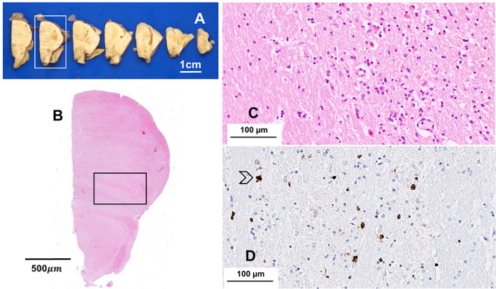
Fig 3.

The axial slices of pons and medulla (A) and the whole mount sections of the pons from the framed slice (B, HE) do not show pathological changes of note; the pons shows increased widespread cellularity secondary to infiltration of atypical cells (C, HE; framed areas in the whole mount); tumor cells often express Ki‐67 indicating proliferation; a mitosis is present in this field (arrow) (D, immunoperoxidase). Scale bars: 1 cm (A), 500 μm (B), 100 μm (C, D).
