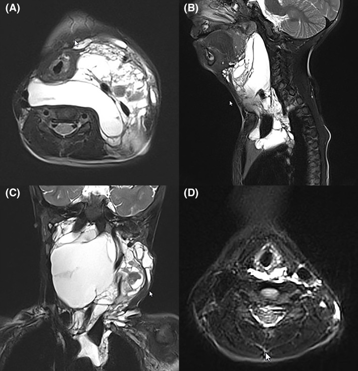
FIGURE 4.

Same child as in Figure 3. A (axial), B (sagittal) and C (coronal) preoperative MRI at 2 years of age. The mixed lymphatic malformation insinuates between cervical plans and causing moderate–severe degree of extrinsic airway obstruction and mediastinal extension. (D) Axial MRI at 7 years of age, following surgery and intralesional sclerotherapy with Bleomycin
