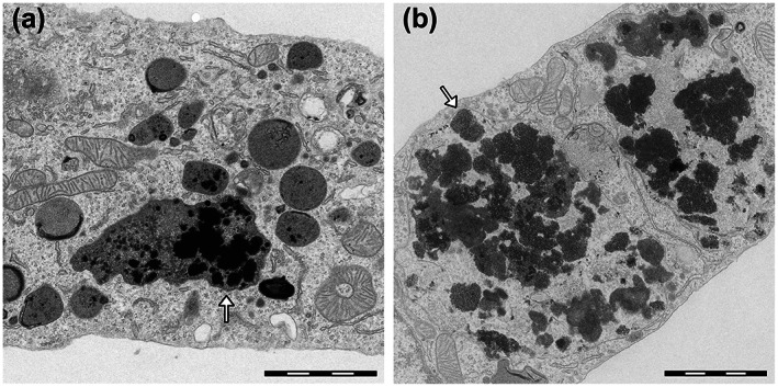
FIGURE 2.

Transmission electron microscopy images of rat microglia cultures exposed to synthetic NM models (a) and human NM pigment isolated from SN (b). Both synthetic and natural NM appear in microscopy images as black and electron‐dense pigments (arrows). The synthetic NM used in this in vitro model has a composition similar to that of the natural pigment, with a melanin‐protein core obtained by melanization of fibrillated β‐lactoglobulin with the presence of iron. Both compounds were actively phagocytosed by microglia cells and engulfed inside specialized phagosomes (arrows) for future breakdown and degradation of the pigments. During this process, this synthetic NM, as well as similar analogues with minor differences in their constituents, were able to promote microglia activation that was assessed by analysis of activation of typical proinflammatory markers in a manner that was similar to that of human NM. Scale bar = 2 μm. Reprinted (adapted) with permission from ref. 15 Copyright 2017 American Chemical Society
