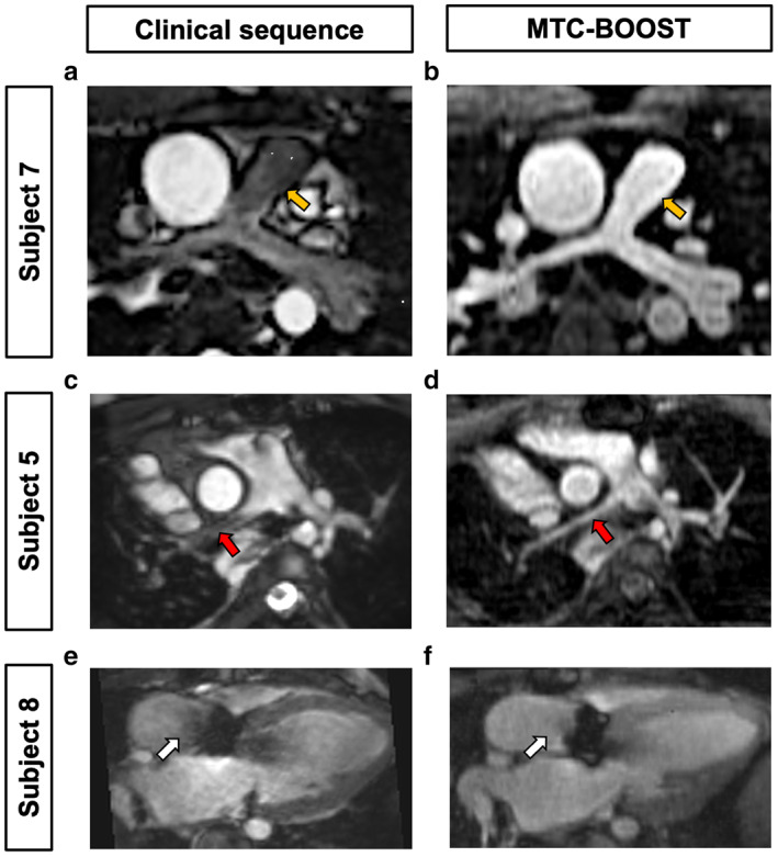
FIGURE 4.

MTC‐BOOST shows resistance to flow‐mediated degradation of luminal signal in comparison to conventional T2prep‐3DWH imaging. Subject 7 has hypoplastic pulmonary arteries (PA) with reduced luminal signal of the main pulmonary artery (yellow arrows) and branch PA vessels using the standard T2prep‐3DWH sequence (a). In contrast, MTC‐BOOST results in clear delineation of the main PA (b). Subject 5 has scimitar syndrome with PA hypoplasia. There is reduced luminal signal in the right PA (red arrow, c) that is mitigated by MTC‐BOOST (d). Subject 8 has prosthetic aortic valve stenosis. There is nonuniform luminal signal in the proximal ascending aorta using the T2prep‐3DWH sequence (white arrow, e) which is not present in MTC‐BOOST images (f).
