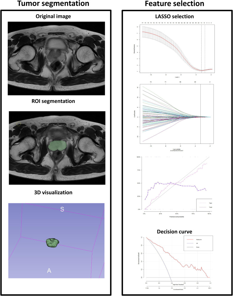
Figure 3.
Workflow of radiomics analysis. The left figure shows the outline of the whole prostate and volume of interest; The right figure shows the method and characteristic coefficient convergence diagram of cross-validation with 10 folds in the Least absolute shrinkage and selection operator (LASSO). By adjusting different parameters λ, the binomial deviation of the implementation model is minimal, to screen out the best feature set. Vertical dotted line indicates the best Log(λ) corresponding to the value λ value. The radiomics workflow started with 3-dimensional segmentation of the whole prostate in magnetic resonance perfusion-weighted imaging (MR-PWI) images. After segmentation, radiomic features including shape, intensity, and texture were extracted with or without wavelet filter of the images. LASSO with 10-fold cross validation was used for the radiomic feature selection. Next, radiomics signature was built with the logistic regression model/combined model, and receiver operating characteristic (ROC) curve was plotted.

