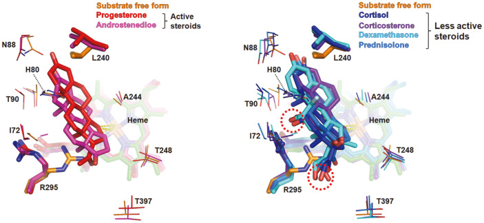
Fig. 5. Overlay of steroids as obtained by superposition onto BaCYP106A6.
The active (left) and less active steroids (right) occupy the substrate-binding pocket depicted in red and blue series colors. Energy-minimized structures and heme molecules are colored the same for each steroid. The hydroxyl groups at 11β and 17β of the less active steroids, marked with red dotted circles, are placed near an Arg295.

