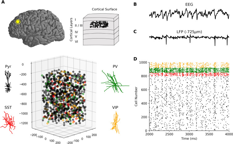
Fig 1. Simulating neuronal spiking and EEG signals from human cortical microcircuits.
(a) Detailed models of human cortical microcircuits, showing somatic placement of 1000 neurons in a 500x500x950 μm3 volume along layer 2/3 (250–1200 μm below pia) and reconstructed morphologies used in the neuron models. (b–d) Temporally aligned multi-scale simulated signals: EEG (b) from scalp electrode directly above the microcircuit; LFP signal (c) recorded at the middle of L2/3 (depth of -725 μm); Raster plot of spiking in different neurons in the microcircuit (d), color-coded according to neuron type. Neurons received background excitatory inputs to generate intrinsic circuit activity.

