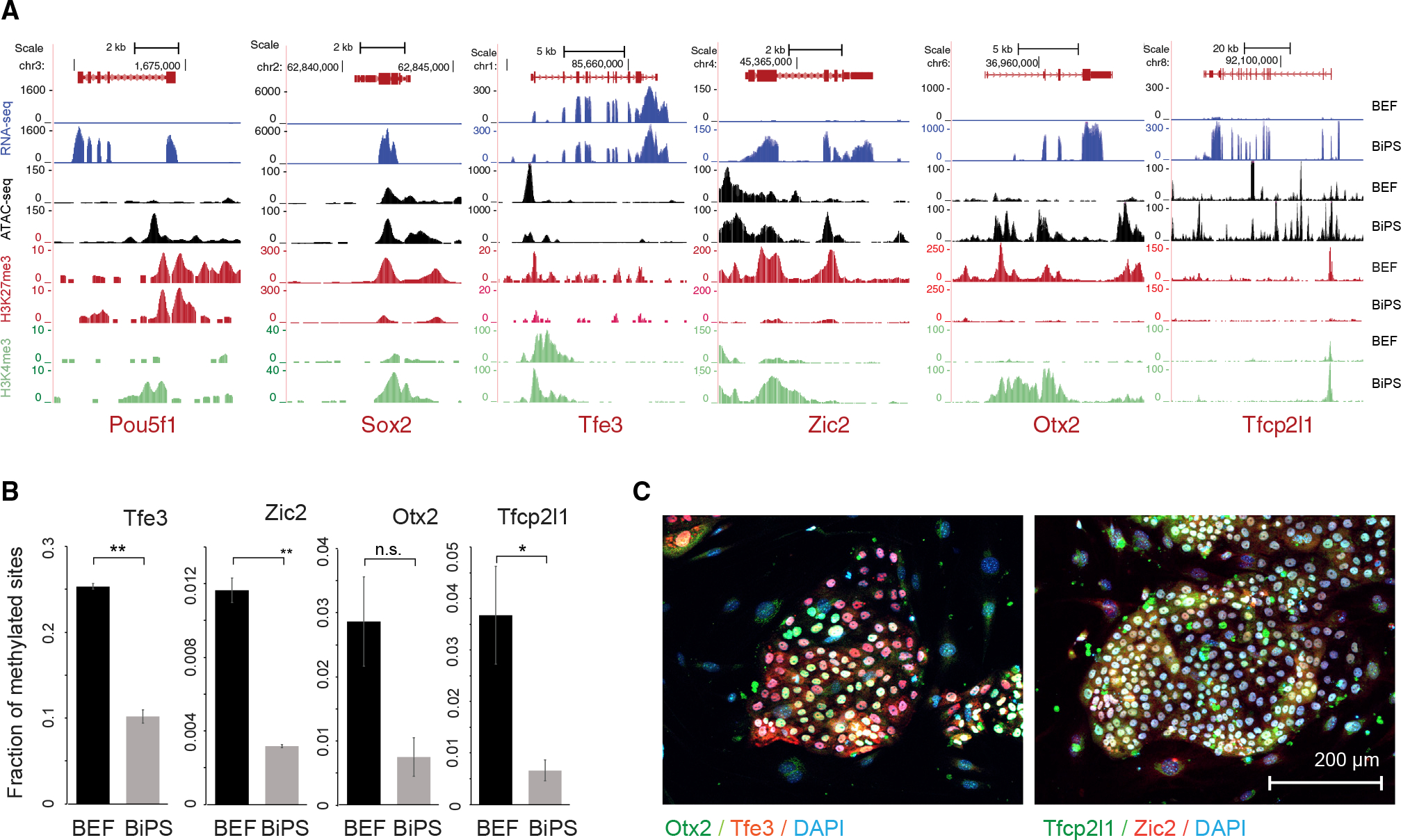
Figure 2. Characteristics of pluripotency markers in pluripotent stem cells generated from Rhinolophus ferrumequinum fibroblasts.

(A) Sequencing tracks showing expression, ATAC-seq signal, histone H3K27 trimethylation (H3K27me3), and histone H3K4 trimethylation (H3K4me3) status of pluripotency markers Oct4 and Sox2 in bat embryonic fibroblasts (BEF) or induced pluripotent stem cells (BiPS).
(B) Fraction of methylated sites in promoters of pluripotency genes that did show promoter methylation. Data are shown as mean ± SD of two replicates; p values were determined by t test: p = 0.0015, 0.0031, 0059, and 0.0481 from left to right. n.s., not significant. Note that we did not detect methylation in the promoters of Nanog, Pou5f1, or Sox2, which might be related to under-annotation of the R. ferrumequinum genome at present.
(C) Immunofluorescence images of bat pluripotent stem cells after staining of markers of naive (Tfe3 and Tfcp2l1) or primed pluripotency (Zic2 and Otx2). See also Table S1.
