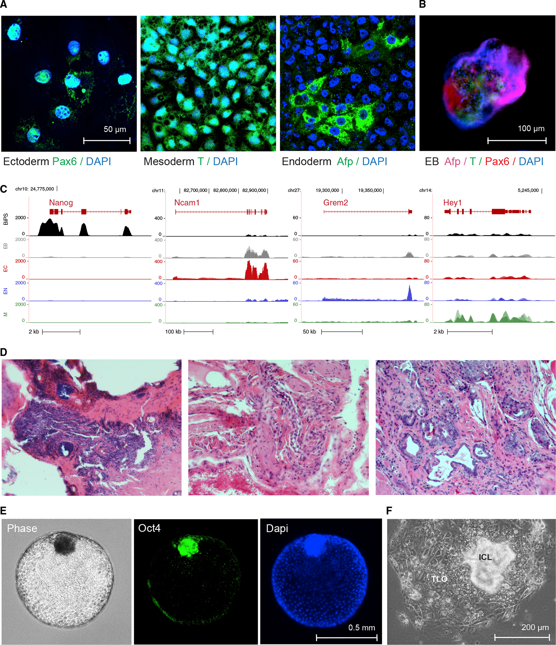
Figure 3. Differentiation potential of R. ferrumequinum bat pluripotent stem cells.

(A) Immunofluorescence microscopy images after staining with antibodies detecting the expression of lineage-specific markers Pax6, Afp, or brachyury (T) following specific directed differentiation into ectoderm, endoderm, or mesoderm, respectively.
(B) Immunofluorescence images of embryonic bodies (EB) that formed after 3D-differentiation of BiPS cells and were stained with antibodies to detect markers specific to all three germ layers as in (A).
(C) RNA-seq signals of selected lineage-specific marker genes in BiPS cells that underwent monolayer differentiation as in (A) or embryonic body differentiation as in (B). Shown is one representative sequencing track (n = 3) per condition. EB, embryonic body differentiation, EC, human ectoderm differentiation protocol; EN, human endoderm differentiation protocol; M, human mesoderm differentiation protocol.
(D) Microscopic images of hematoxylin-eosin-stained sections of tumor tissue after injection of BiPS cells into immunocompromised mice exhibiting ectodermal (left), mesodermal (middle), and endodermal (right) features.
(E) Images of floating blastoids that were obtained from BiPS cells after exposure to Bmp4 to capture their morphology by phase-contrast microscopy (left) and to detect Oct4 expression in inner-cell mass-like cell clusters after immunofluorescence staining (middle, right).
(F) Phase-contrast microscopy image of a typical blastocyst-outgrowth-like cell cluster that formed after the attachment of blastoids to the cell culture vessel surface during Bmp4-induced differentiation as in (E). ICL, inner cell mass-like; TLO, trophoblast-like outgrowth.
