
Figure 5.
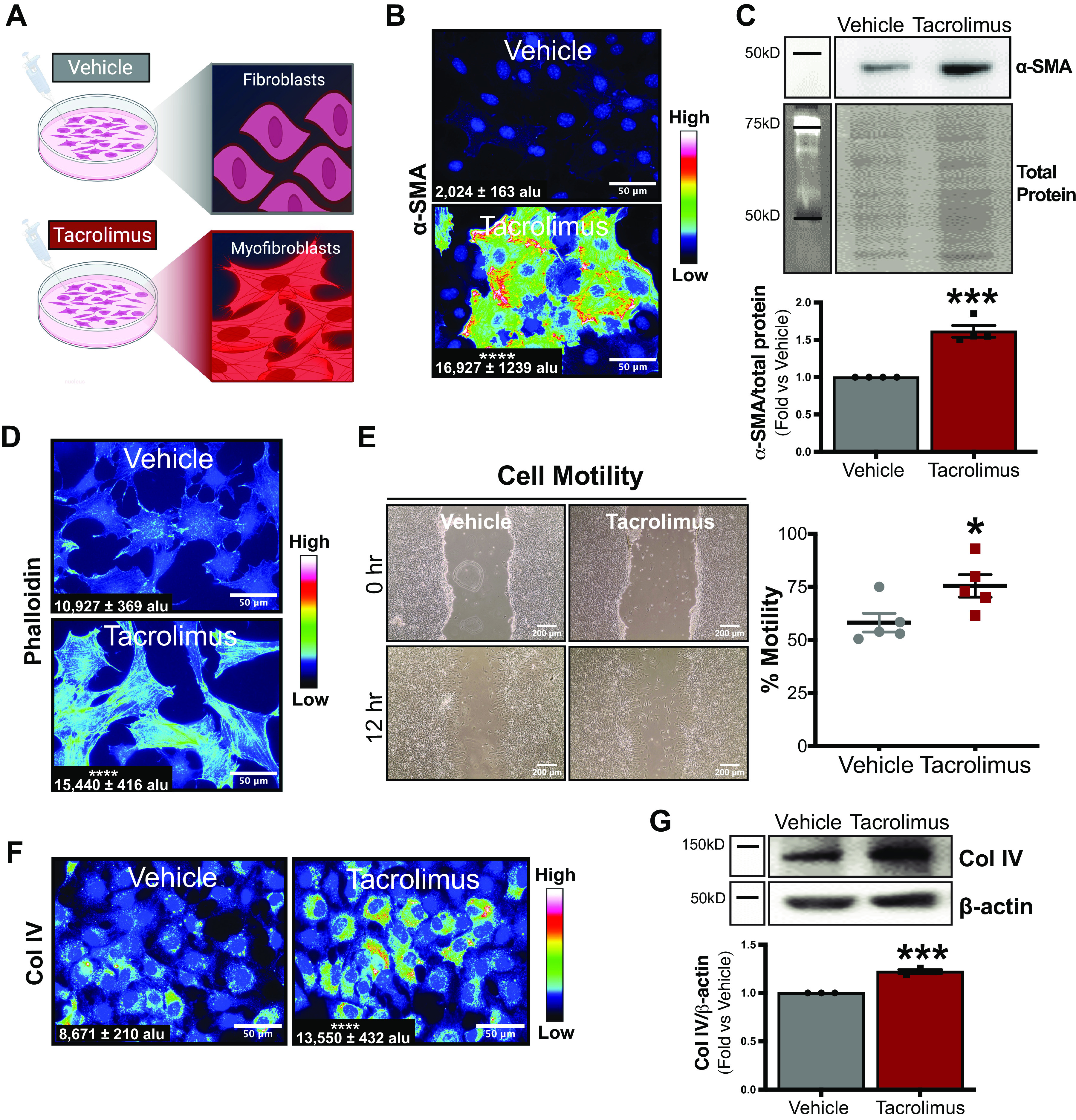
Tacrolimus promotes fibroblast-to-myofibroblast transition (FMT) and extracellular matrix production. To investigate fibroblasts as a source of tacrolimus-induced renal fibrosis, renal fibroblasts were treated with either tacrolimus (1 nM) or vehicle (0.01% ethanol) for 24 h (A). Myofibroblast marker α-smooth muscle actin (α-SMA) protein was visualized via immunofluorescence (B) and quantified with CellProfiler software [expressed as arbitrary light units (alu)]. Western blot (C) also quantified α-SMA expression. To further assess FMT, actin stress fiber content was visualized by immunofluorescent staining of phalloidin (D) and quantified with CellProfiler software (expressed as alu). To evaluate the effect of tacrolimus on fibroblast motility, scratch test assays were conducted and cell motility rates (in %) were calculated (E). Extracellular matrix production was assessed by both immunofluorescent detection and quantification of collagen type IV (Col IV) protein (with CellProfiler software, expressed as alu) (F). Western blot analysis further quantified Col IV expression (G). Representative images of at least 3 or 4 independent studies are shown. Values are means ± SE. Statistical tests were conducted using a t test to detect differences between experimental groups. *P < 0.05, ***P < 0.0005, and ****P < 0.00005 vs. vehicle.