Figure 5.
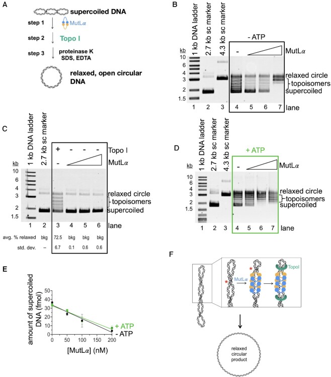
MutLα stimulates DNA relaxation by a topoisomerase in vitro. (A) Schematic depicting topoisomerase assay. Supercoiled pUC18 DNA was incubated with MutLα (step 1) followed by addition of E. coli Topoisomerase I (step 2) which converts supercoiled DNA into relaxed, open circular DNA. The protein component of reactions is degraded and the DNA end products are analyzed by native, ethidium bromide stained agarose gel. (B) Agarose gel depicting DNA relaxation by Topo I and MutLα in the absence of ATP. Lane 1 contains 1 kb plus DNA marker and lanes 2 and 3 contain 2.7 and 4.3 kb supercoiled (sc) DNA markers respectively. Lane 4 is a negative control without MutLα and lanes 5–7 contain a titration of MutLα (50, 100 and 200 nM). (C) Agarose gel depicting MutLα activities in this assay in the absence of Topo1. Lane 3 is a positive control with Topo1. In lanes 4–6, MutLα was titrated at 50, 100 and 200 nM final concentrations. Number of replicates is 3. (D) Experiment and gel are identical to those in panel B, but in the presence of 0.25 mM ATP. (E) Plot for gels in B and D showing amount of supercoiled DNA versus MutLα concentration. Black circles are conditions without ATP and green squares are conditions with ATP. Experiment was performed in triplicate. Data was fit to a simple linear regression curve with error bars representing the standard deviation between experiments. (F) Model for MutLα activities in this assay. Red asterisk indicates the repositioning of the helical crossover.

