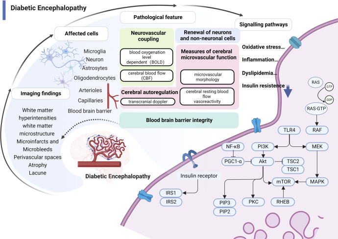
Fig. 3.
Pathology and molecular mechanisms of DE. (1) Structural brain changes from MRI studies in diabetes are the primary diagnostic basis of DE, including microinfarcts and microbleeds, perivascular spaces, white matter hyperintensities, white matter microstructure, lacunes, and atrophy; (2) Pathologies related to imaging findings include blood-brain barrier permeability, perfusion defects, hypoxia, and increased angiogenesis that can involve brain microvessels, multiple nerve cells, and the blood-brain barrier; (3) Microvascular dysfunction manifested by impaired neurovascular coupling and impaired neuronal function. Neurovascular coupling links transient local neural activity to subsequently increased blood flow; (4) The molecular pathways of hyperglycemic damage to brain microvasculature are closely related to oxidative stress, inflammation, abnormal lipid metabolism, and insulin resistance. RAS rat sarcoma protein, GTP guanosine triphosphate, GDP guanosine diphosphate, TLR4 Toll-like receptor 4, MEK mitogen-activated protein kinase, PI3K phosphoinositide 3-kinase, Akt protein kinase B, TSC tuberous sclerosis complex, MAPK mitogen-activated protein kinases, mTOR mammalian target of rapamycin, RHEB Ras homolog protein enriched in brain, PKC protein kinase C, PGC1-α PPAR-gamma co-activator-1 alpha, PIP2,3 phosphatidylinositol bisphosphate2, 3, IRS-1,2 insulin receptor substrate1, 2, NFκB nuclear factor kappa-B

