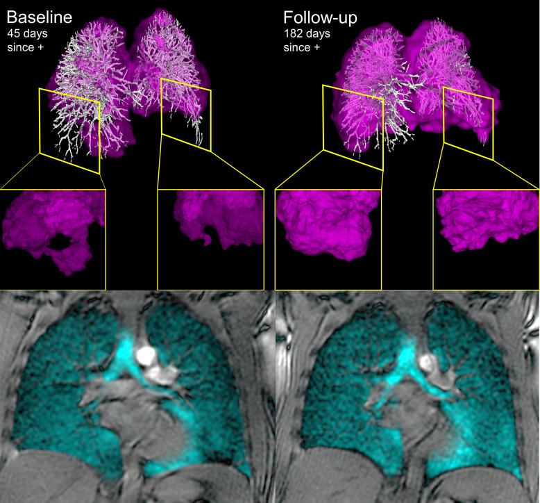
Figure 2.
129Xe MRI and coregistered pulmonary vascular tree CT at baseline and follow-up. Left and right panels show 129Xe MRI RBC map (pink) coregistered with CT pulmonary vascular tree (white) and bottom panels show 129Xe ventilation images (cyan) for a previously healthy participant hospitalised with COVID-19 symptoms and pulmonary embolism. At baseline, 45 days post-COVID-19 positive test, RBC:TP ratio was abnormally low (0.37) and insets provide examples of RBC map defects. At follow-up the RBC:TP ratio improved (0.54) as did the lower lobe red blood cell map defects shown in the right panel inset. DLCO (baseline=93%pred, follow-up=110%pred) and total SGRQ score. (baseline=23, follow-up=5) also improved at follow-up. DLCO, diffusing capacity of the lung for carbon monoxide; RBC, red blood cell; SGRQ, St George’s Respiratory Questionnaire.

