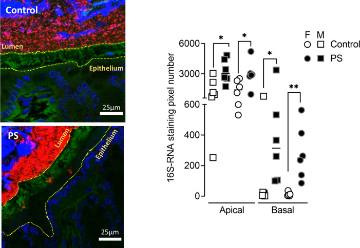
Figure 3.
PS alters gut microbiota spatial organisation in adulthood. Bacteria were labelled with the universal probe Eub338 (red); wheat germ agglutinin-Fluorescein-5-isothiocyanate (FITC) was used to stain the polysaccharide-rich mucus layer (green); and the epithelial cell nucleus was stained with 4',6-diamidino-2-phénylindole (DAPI; blue). Bacteria penetration into the mucus was measured in both M (square) and F (circle) control (white symbols) and PS (black symbols) mice by image processing on Fiji by quantifying the number of 16S RNA-labelled pixel between the edge of the lumen and the middle of the mucus (apical) and between the middle of the mucus and the edge of the epithelium (basal). The results are expressed as scatter dot plot with the mean. Statistical analysis was performed using Mann-Whitney test. * P<0.05, **P<0.01, significantly different from the control group. (n=12 mice/group; four images/mice, two independent experiments). F, female; M, male; PS, prenatal stress.

