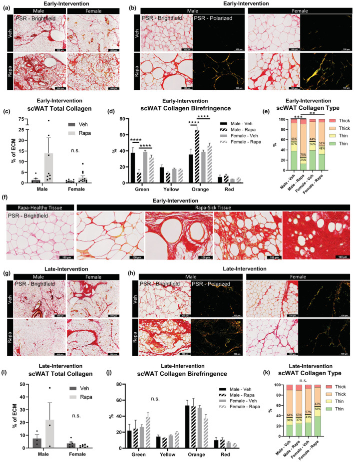
FIGURE 8.

Rapamycin treatment started early in life increased scWAT fibrosis. PSR staining of scWAT from early‐intervention (a–f) versus late‐Intervention (g–k) of rapamycin (42 ppm) treated male and female HET3 mice imaged at 4× objective magnification (a,g). Five representative images of tissue parenchyma were captured separately with bright‐field and polarized light at 20× objective magnification per tissue per animal (b,h). Total collagen was measured as a ratio of birefringent collagen to total PSR staining (c,i). Changes in specific hues of collagen birefringence (d,j). Contribution of collagen fiber thickness determined by hue (e,k). Thin collagen (green and yellow); thick collagen (orange and yellow). N = 3–11. Two‐way ANOVA with Tukey's correction for multiple comparisons (c,d,i,j). One‐way ANOVA with Tukey's correction for multiple comparisons (e,k). All error bars are SEMs. n.s. p > 0.05, *p < 0.05, **p < 0.01, ***p < 0.001, ****p < 0.0001
