Figure 1.
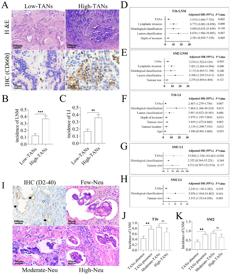
TANs promote tumor invasion in gastric cancer. (A) TANs were detected in the tumor stroma of nearly all T1b gastric cancer tissues by H & E staining and IHC. In low-TANs group, TANs were rare and occasionally present, and the enlarged frame showed few neutrophils with CD66b staining. In high-TANs group, more than 10 neutrophils per HPF were detected, and the enlarged frame showed multiple neutrophils with CD66b staining. (B) The incidence of LNM in high-TANs group was much higher than that in low-TANs group (P = 0.000). (C) The incidence of lymphatic invasion (LI) in high-TANs group was much higher than that in low-TANs group (P = 0.001). (D) The multivariate analysis indicated that Lauren classification, lymphatic invasion and high TANs are the independent risk factors for LNM in T1b tumors. (E) Lauren classification, lymphatic invasion and high TANs were the independent factors for LNM in SM2 tumors. (F, G, H) TANs served as one of the independent risk factors for lymphatic invasion (LI) in T1b, SM1 and SM2 tumors respectively. (I) Neutrophils could be detected in about half of lymphatic cancer emboli in T1b tumors. IHC with anti-D2-40 showed the lymphatics (black arrow) in tumor tissues, and blank arrows show the cancer embolus and the neutrophils in cancer emboli. (J, K) The incidence of LNM in patients with neutrophils in lymphatic cancer emboli was significantly increased with regard to that in patients without neutrophils in cancer emboli in either T1b tumors (J) or in SM2 tumors (K). However, the neutrophil numbers in emboli had no influence on LNM (J, K). (**, P < 0.01; ***, P < 0.001) (Neu, neutrophil)

