
FIGURE 2.
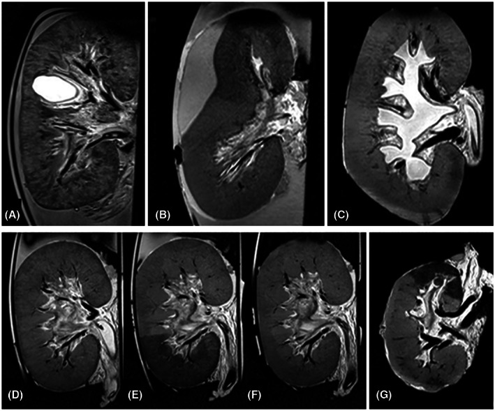
T2‐weighted images of structural abnormalities and ischemia/reperfusion damage. (A) Renal cyst not visible by visual inspection of the kidney's surface. (B) Subcapsular hematoma compressing part of the parenchyma and large intrarenal vessels. (C) Hydronephrosis due to an external obstruction of the ureter cannula. (D–F) Porcine kidney without ischemia, 60 min after induced partial ischemia (segmental area with lower T2 signal) and 30 min after reperfusion (segmental T2 signal almost recovered to its initial intensity). (G) Human discarded kidney (without intervention) with regional perfusion defect only visible on MRI.