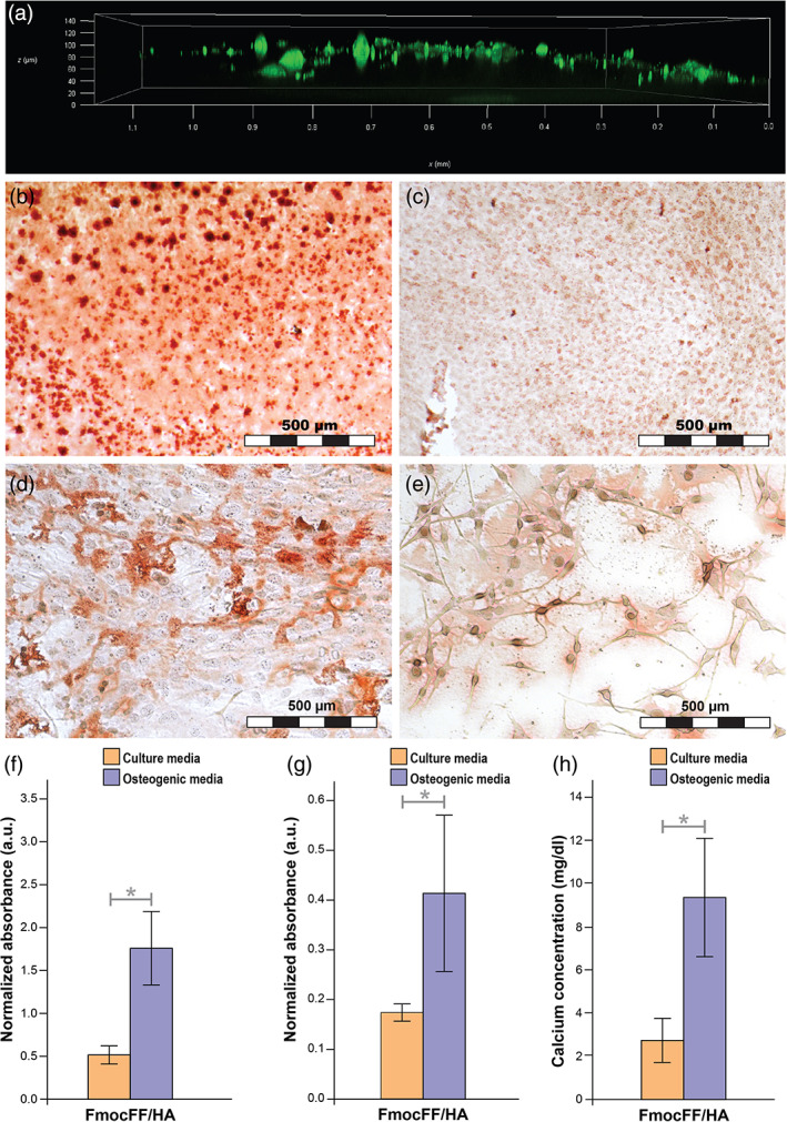
FIGURE 3.

Migration and osteogenic differentiation of MC3T3‐E1 preosteoblasts on the hydrogel (a) migration of MC3T3‐E1 preosteoblasts 1 day after seeding using confocal microscopy. (b) Optic microscope images of MC3T3‐E1 preosteoblasts stained with Alizarin red 14 days after seeding on FmocFF/hyaluronic acid (HA) hydrogel with osteogenic media. (c) Optic microscope images of MC3T3‐E1 preosteoblasts stained with Alizarin red 14 days after seeding on FmocFF/HA hydrogel with culture media. (d) Optic microscope images of MC3T3‐E1 preosteoblasts stained with Alizarin red 14 days after seeding on the plate with osteogenic media. (e) Optic microscope images of MC3T3‐E1 preosteoblasts stained with Alizarin red 14 days after seeding on the plate with culture media. (f) Normalized Alizarin red staining absorbance values 14 days after MC3T3‐E1 osteogenic differentiation with and without osteogenic media. (g) Normalized alkaline phosphatase activity of MC3T3‐E1 preosteoblasts 14 days after seeding with and without osteogenic differentiation. (h) Quantification of calcium content in the supernatant of the FmocFF/HA hydrogel 14 days after seeding with and without osteogenic media. Data analysed using a two‐tailed Student's t‐test. *p < .05, **p < .01
