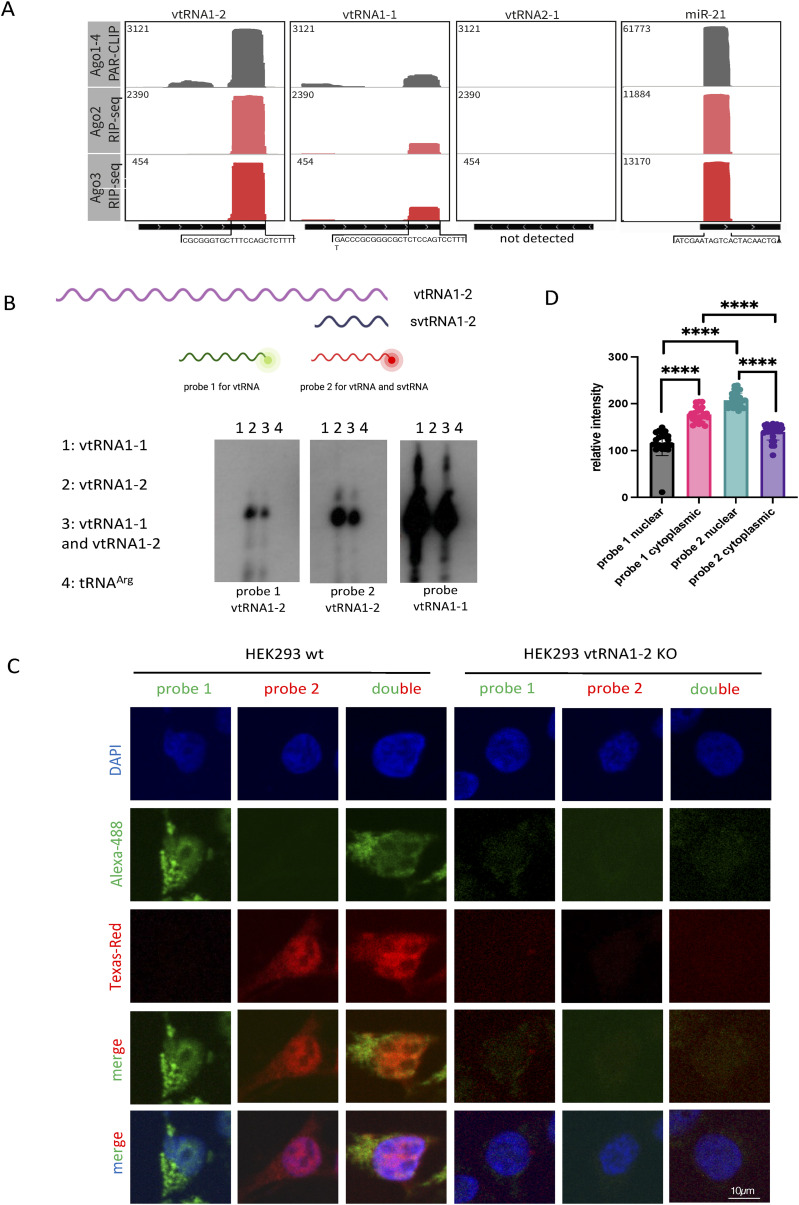
Figure 2. svtRNA1-2 is bound to Argonaute proteins.
(A) Integrative Genomics Viewer tracks of AGO1-4 PAR-CLIP and AGO2/3 RIP-seq show the association of AGO proteins with svtRNA1-2 (data values are shown in the top left corner). miR-21 was used as a positive control. The bottom black line indicates the length of the transcript, and the sequence below represents the interacting fragment. Note: no reads were detected for vtRNA2-1. (B) Diagram illustrating the position of probes 1 and 2 with respect to vtRNA1-2 and svtRNA1-2. Representative Northern blot showing signals for vtRNA1-1 and vtRNA1-2 when incubated with probes 1 and 2 designed for vtRNA1-2. (C) Representative FISH confocal images showing fluorescent signals for probes 1 and 2 in HEK293 WT and vtRNA1-2 KO cells. DAPI was used to stain nuclei. (D) Bar chart showing relative fluorescence intensity levels of probes 1 and 2 in the nucleus and the cytoplasm. Error bar = mean ± SEM; significance was determined using a multiple unpaired t test, ****P ≤ 0.0001.

