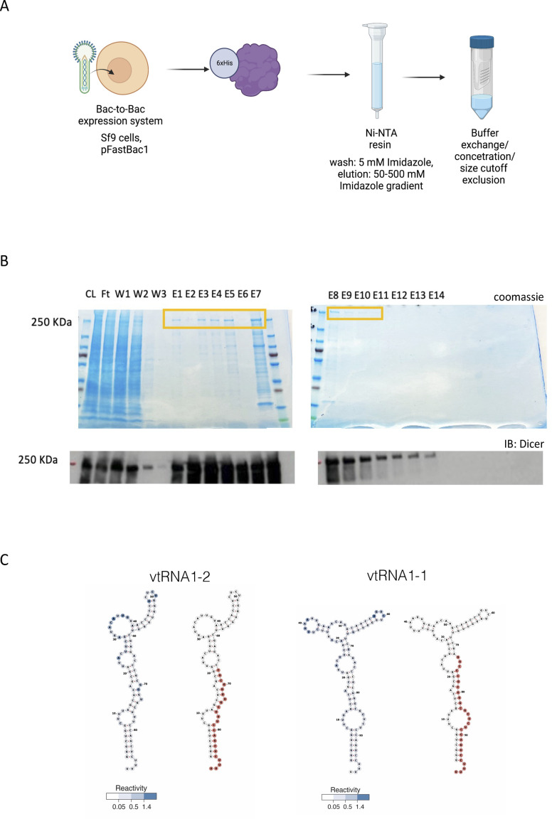
Figure S2. Purification of recombinant Dicer.
(A) Diagram summarising 6xHis-Dicer purification strategy from insect cells. (B) Representative protein gel images showing fractions with a purified Dicer protein stain by Coomassie (upper image) or detected by Western blot using anti-Dicer antibody (lower image) (CL, cell lysate; FT, flowthrough; W, washes; and E, elution). (C) Predicted secondary structures of vtRNA1-1 and vtRNA1-2. Reactivity score is shown in blue.

