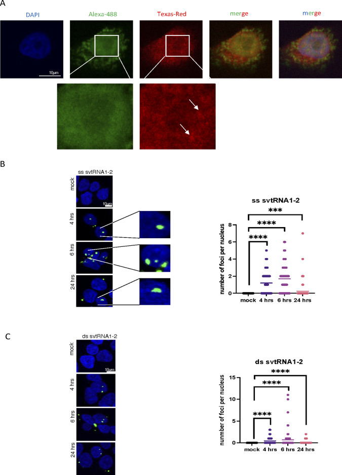
Figure S5. svtRNA1-2 is localised in nucleus.
(A) Representative confocal images showing fluorescence of probes 1 (green) and 2 (red) as in Fig 2, in HeLa cells. Zoomed parts are depicted in white squares, and clusters are indicated by arrows. DAPI was used to stain nuclei. (B) Confocal microscopy images show nuclear localisation of double-stranded (ds) svtRNA1-2 (green) at different time points. DAPI was used to stain nuclei of the cells. Images were quantified using CellProfiler software. Significance was calculated using a t test (****P < 0.0001 and ***P < 0.001). (C) Confocal microscopy images show nuclear localisation of single-stranded (ss) svtRNA1-2 (green) at different time points. DAPI was used to stain the nuclei of the cells. Images were quantified using CellProfiler software. Significance was calculated using a t test (****P < 0.0001 and ***P < 0.001).

