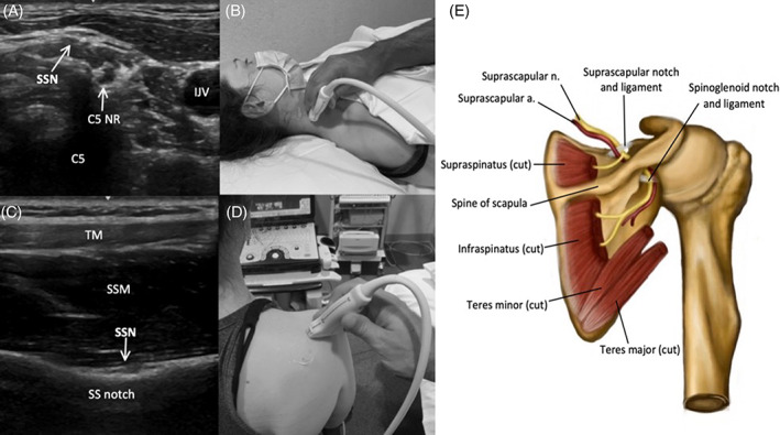
FIGURE 3.

Image 3A demonstrates the SSN in the posterior triangle of the neck, shortly after branching from the C5 nerve root (C5 NR) (C5 = shadow from posterior tubercle of C5 foramen, and Image 3B shows the corresponding probe placement. Image 3C demonstrates the SSN passing through the suprascapular notch (SS notch), deep to the TM and SSM, and Image 3D shows the corresponding probe placement. Image 3E depicts the SSN and relevant anatomy in the posterior scapular region
