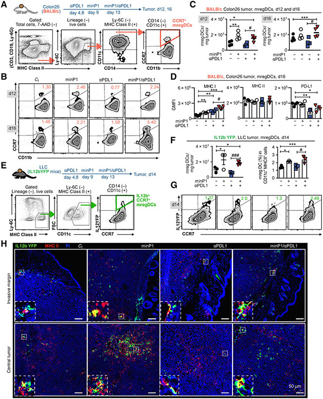
Figure 4.
minP1 increases the number of intratumoral mregDCs at both the invasive margin and central tumor and enhances their MHC class I expression. A, Schema on the preparation of tumor immune cells from Colon26 tumor–bearing BALB/c mice and flow cytometry gating of CCR7+ mregDCs. B and C, The frequency and number of CCR7+ mregDCs in the tumors on days 12 and 16 after tumor inoculation. D, The expression of MHC class I, MHC class II, and PD-L1 on CCR7+ mregDCs on day16. GMFI, geometric MFI. E, Schema on the preparation of tumor immune cells from LLC tumor–bearing IL12b-YFP reporter C57BL/6 mice and flow cytometry gating of IL12b+CCR7+ mregDCs. F and G, Frequency and number of the IL12b+CCR7+ mregDCs in tumors from LLC tumor–bearing IL12b-YFP reporter C57BL/6 mice treated with combination treatment on day 14 after tumor inoculation. H, The number and localization of IL12b+CCR7+ mregDCs in LLC tumor sections identified by coexpression of IL12b-YFP (green) and MHC class II (red), with a 200 μm scale bar. Bottom panels indicate zoomed and merged images of costained mregDCs. Each result is representative of three independent experiments with at least four mice/group. Data are presented as mean ± SEM. *, P < 0.05; **, P < 0.01; ***, P < 0.001 using Dunnett post hoc test (compared with control); #, P < 0.05; ##, P < 0.01; ###, P < 0.001 using Student t test (comparing with minP1/anti–PD-L1–treated and anti–PD-L1–treated groups).

