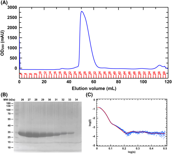
Fig. 2.

Purification and biophysical characterization of PaNADK. (A) Elugram from the gel filtration column. OD at 280 nm is shown as blue line. Fraction numbers are shown in red. (B) Verification of the purity of the recombinant PaNADK by SDS/PAGE gel. The numbers of the fractions obtained from the GF are indicated at the top of the gel. (C) Scattering profiles obtained by SAXS on PaNADK and fitting with a tetrameric model. The fitting of homo‐tetrameric protein solution scattering obtained from X‐ray crystallography (red line) to the experimental scattering obtained from SAXS experiments (blue dots).
