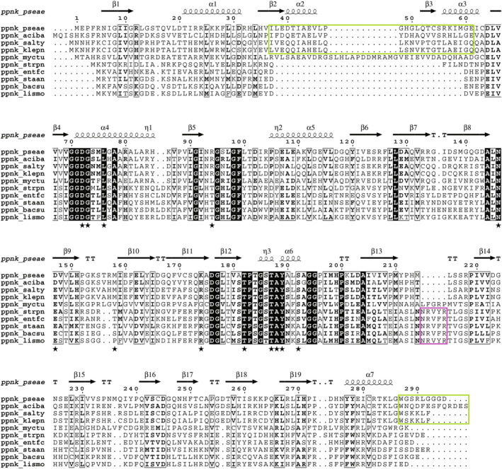
Fig. 5.

Sequence alignment of bacterial NADKs. Sequences of bacterial NADKs were aligned and classified from top to bottom as follows: NADKs of Gram‐negative bacteria (Pseudomonas aeruginosa, Acinetobacter baumannii, Salmonella typhimurium and Klebsiella pneumoniae), of Mycobacterium tuberculosis and of Gram‐positive bacteria (Streptococcus pneumoniae, Enterococcus faecium, Staphylococcus aureus, Bacillus subtilis and Listeria monocytogenes). Conserved residues are highlighted by black boxes, and conservative substitutions are shown in bold. Extensions specific to Gram‐negative and Gram‐positive bacteria are highlighted by green and pink boxes, respectively. Residues important for NADP+ binding are indicated by a black star symbol. Secondary structures in PaNADK are shown above the alignment. The figure was drawn using ESPript [44].
