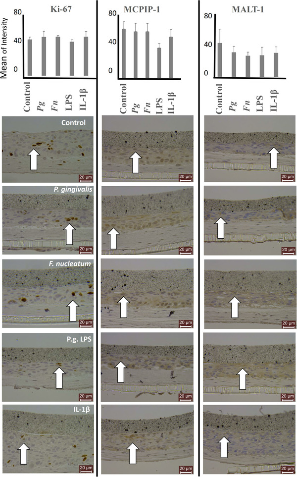
FIGURE 6.

MCPIP‐1 and MALT‐1 protein expression levels of organotypic oral mucosal models after incubation with P. gingivalis, F. nucleatum, P. gingivalis LPS, and IL‐1β. Cellular proliferation is detected with Ki‐67 stainings. Bars indicate mean values and standard deviations. White arrows indicate representative positively stained cells
