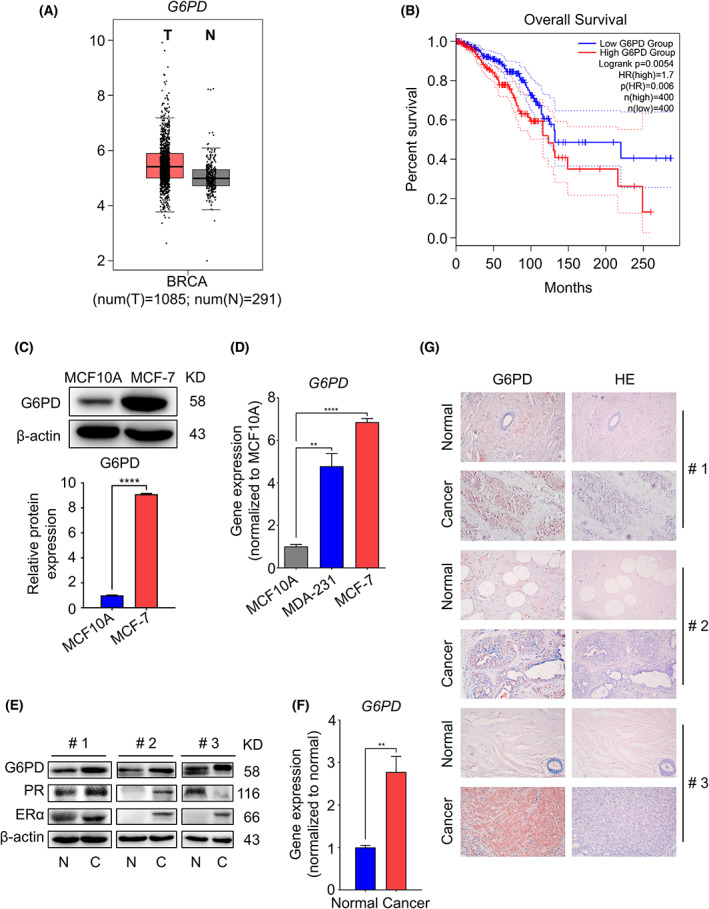
Fig. 1.

Identification of abnormally activated G6PD in breast cancer. (A) Gene expression of G6PD in normal people (n = 291) and breast cancer patients (n = 1085) was analysed by the StarBase 3.0 website. (B) The relationship between G6PD expression and the survival rate in human samples [n (high) = 400, n (low) = 400] was analysed by the StarBase 3.0 website. (C) Immunoblot analyses of and G6PD in MCF‐7 cells compared to MCF‐10A cells. (D) q‐PCR analysis of the transcription levels of G6PD in MDA‐231 and MCF‐7 cells compared to MCF‐10A cells. (E) Immunoblot analyses of G6PD, PR, and ERα protein in breast cancer patients compared to normal people (N: Normal, C: Cancer). (F) Transcription levels of G6PD were measured using q‐PCR on mRNA prepared from breast cancer patients and normal breast samples. (G) IHC staining detected G6PD in breast cancer tissue and paracarcinoma tissue in humans. 200× magnification. **P < 0.01, ****P < 0.001.
