
FIGURE 3.
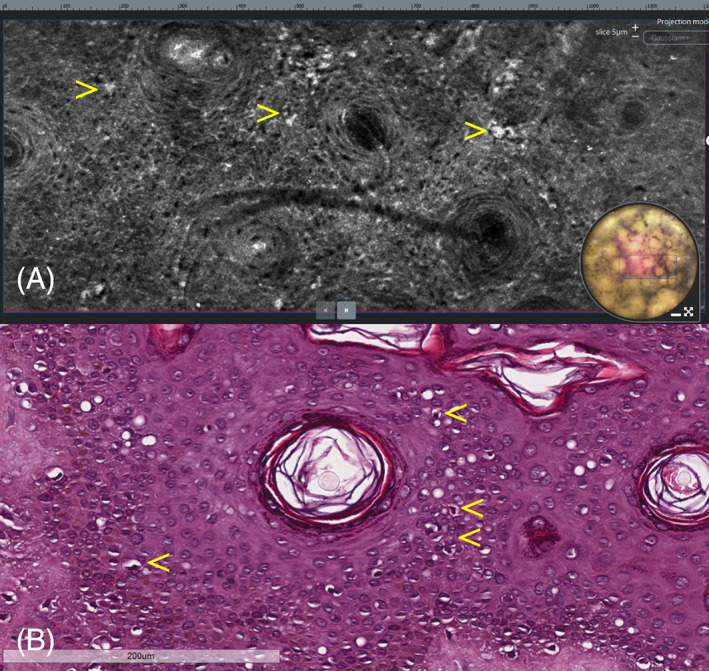
Lentigo maligna. (A) Horizontal LC‐OCT at the level of the stratum spinosum showing bright atypical, roundish cells with evident nuclei (arrows), causing architectural disarray. (B) Horizontal histopathologic section at the same level showing intraepidermal pagetoid spread of scattered neoplastic melanocytes (arrows). H&E: original magnification, ×150