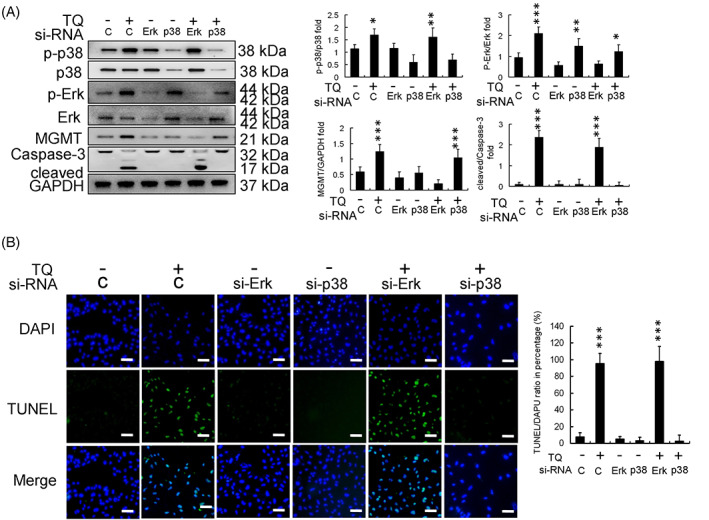
FIGURE 4.

Effect of phosphorylated‐extracellular signal‐regulated kinase (p‐ERK) and p38 protein silencing in thymoquinone (TQ)‐treated U251R cells. (A) Protein expression and calibration. (B) DAPI‐TUNEL staining assay (scale bar in each image: 100 μm) showed that si‐p38 inhibited U251R cell apoptosis and si‐ERK enhanced U251R cell apoptosis in the background of 24 h TQ (50 μM) treatment in U251R cells. (*p < .05, **p < .01, ***p < .001, compared with control). DAPI, 4,6‐diamidino‐2‐phenylindole; ERK, extracellular signal‐regulated kinase; GAPDH, glyceraldehyde 3‐phosphate dehydrogenase; MGMT, methyl guanine methyltransferase; siRNA, small interfering ribonucleic acid; TUNEL, terminal deoxynucleotidyl transferase dUTP‐mediated nick‐end labeling
