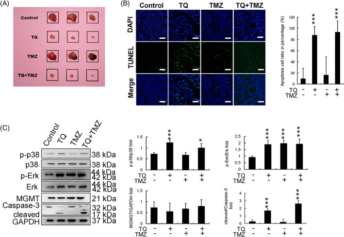
FIGURE 5.

Evaluation of the anticancer effect of thymoquinone (TQ) in vivo. (A) Tumors arising from U251R cells in a mouse xenograft model (for each treatment group); square size is 20 mm × 20 mm. Tumor volumes are reduced in the TQ and TQ + TMZ treatment groups. (B) DAPI‐TUNEL staining assay (scale bar in each image: 100 μm). Apoptotic cell percentage was increased in the TQ and TQ + TMZ treatment groups. (C) Protein expression and calibration in tissue samples. TQ enhanced the levels of p‐p38 and regulated downstream caspase‐3 activation. (*p < .05, **p < .01, ***p < .001, compared with control). DAPI, 4,6‐diamidino‐2‐phenylindole; ERK, extracellular signal‐regulated kinase; GAPDH, glyceraldehyde 3‐phosphate dehydrogenase; MGMT, methyl guanine methyltransferase; p‐ERK, phosphorylated extracellular signal‐regulated kinase; TMZ, temozolomide; TUNEL, terminal deoxynucleotidyl transferase dUTP‐mediated nick‐end labeling
