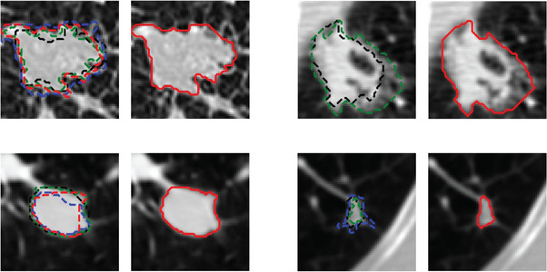
FIGURE 2.

Examples of radiologists' annotations on four different nodules showing the variabilities among radiologists in manual outlining of nodules (left, dashed contours with different colors). The 50% consensus consolidation of radiologists' annotations for each nodule was used as the reference standard (right, red solid contour)
