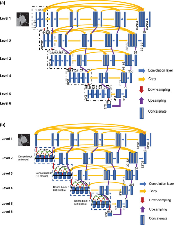
FIGURE 3.

The architectures of our two asymmetric U‐shaped deep learning backbone networks for lung nodule segmentation in CT images. (a) VGG19‐based encoder path (Shallow U‐DL) (upper), and (b) deep DenseNet‐based encoder path (Deep U‐DL) (bottom). The size of each feature map is shown at the lower‐left edge of the box. The arrows of different colors represent different operations.
