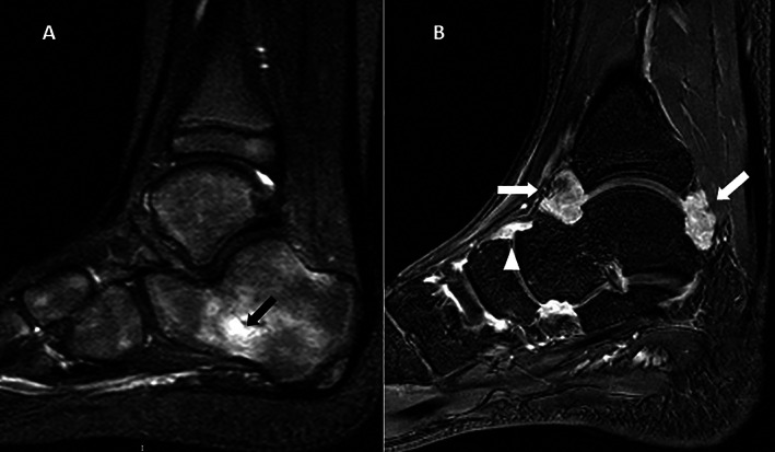
Figure 1.

Contrast‐enhanced magnetic resonance imaging of the ankle obtained from 2 patients with juvenile idiopathic arthritis in clinical remission. A, Sagittal T2‐weighted sequence showing bone marrow edema within the calcaneus (arrow). B, Sagittal T1‐weighted gadolinium contrast‐enhanced sequence showing synovial enhancement in the tibiotalar (arrows) and talo‐navicular (arrowhead) joints.
