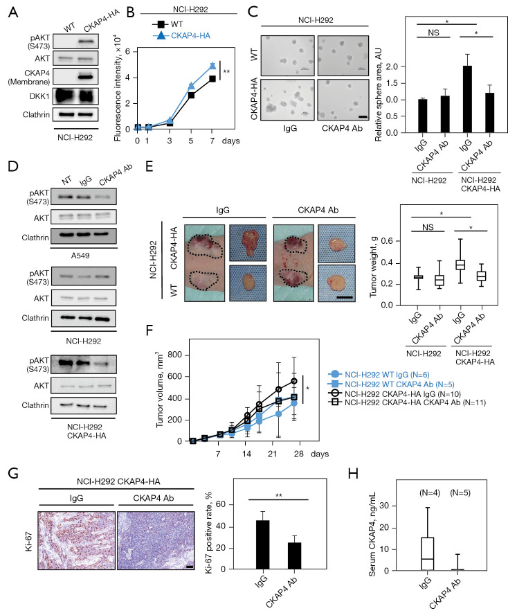
Figure 4.
Anti-CKAP4 antibody inhibits NSCLC growth in vitro and in vivo. (A) Lysates of WT NCI-H292 or NCI-H292/CKAP4-HA cell were probed with indicated antibodies. (B) WT NCI-H292 or NCI-H292/CKAP4-HA cells were subjected to a 2D cell proliferation assay using CyQUANT NF. CyQUANT NF dye fluorescence intensity was measured on each day. (C) Left panels: WT NCI-H292 or NCI-H292/CKAP4-HA cells treated with 20 µg/mL anti-CKAP4 or control IgG antibodies were cultured for 5 days in 3D Matrigel, and were observed with a phase contrast microscope. Right panel: the total area of spheres/field (n=5 fields) were quantified. (D) WT-A549, WT-NCI-H292, or NCI-H292/CKAP4-HA cells treated with 20 µg/mL anti-CKAP4 or control IgG antibodies for 4 h. (E) Left panels: WT-NCI-H292 or NCI-H292/CKAP4-HA cells were subcutaneously implanted into immunodeficient mice. An anti-CKAP4 antibody or control IgG (200 µg/body) was injected into the intraperitoneal cavity twice a week. A representative mouse (left) and extirpated xenograft tumors (right) are shown. Dashed lines show xenograft tumor outlines. Right panels: extirpated xenograft tumor weights were measured. (F) Xenograft tumor volumes in (E) were measured at indicated times. (G) Left panels: sections prepared from xenograft tumors in (E) were stained with hematoxylin and an anti-Ki-67 antibody. Right panel: percentage of Ki-67-positive cells are expressed (n=5 fields). (H) CKAP4 serum levels from mice with xenograft tumors in (E) on the final day were measured using sandwich ELISA. Results are shown as the mean ± SD (B, C, F, and G). Results are indicated by a box plot. The median is represented with a line, the box represents the 25th–75th percentile, and error bars show the 5th–95th percentile (E and H). *, P<0.05; **, P<0.01 (Student’s t-test) (C and G); *, P<0.05; **, P<0.01 (Wilcoxon’s rank-sum test) (B, E and F). Scale bars, 100 µm (C and G); 10 mm (E). WT, wild-type; NS, not significant; AKT, Ak strain transforming; NT, no treatment; Ab, antibody; NSCLC, non-small cell lung cancer; ELISA, enzyme-linked immunosorbent assay; SD, standard deviation; IgG, immunoglobulin G.

