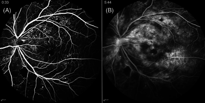
FIGURE 10.

Widefield (WF) fundus fluorescein angiography (FFA) of a left eye with proliferative diabetic retinopathy. Numerous microaneurysms (MAs), neovascularization elsewhere (NVE), neovascularization of the disc (NVD), capillary nonperfusion (CNP) and an irregular foveal avascular zone (FAZ) are observed in early phase of FFA (A). In the late phase (B), the leakage from MAs, NVE and NVD is profound. The images were obtained using the Spectralis HRA + OCT (Heidelberg Engineering, Heidelberg, Germany)
