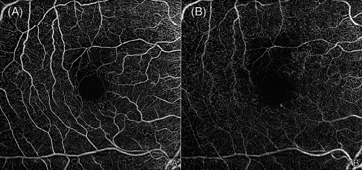
FIGURE 11.

6 × 6 mm en face optical coherence tomography angiography (OCTA) images of a right eye with mild nonproliferative diabetic retinopathy (NPDR). Microaneurysms (MAs), an enlarged and irregular foveal avascular zone, and some small regions of capillary dropout area are observed in the superficial (A) and deep (B) capillary plexuses. Of note, projection artefacts from the superficial vessels are seen on the deep plexus image (B) as the projection removal was not employed. The images were obtained using the PLEX Elite 9000 (Carl Zeiss Meditec, Dublin, CA)
