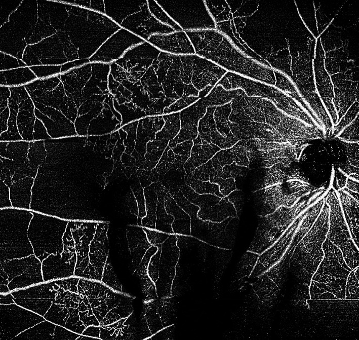
FIGURE 12.

12 × 12 mm en face optical coherence tomography angiography (OCTA) of a right eye with proliferative diabetic retinopathy (PDR). Microaneurysms (MAs), intraretinal microvascular anomalies (IRMAs) and numerous areas of capillary dropout are observed in superficial plexus of right eye. Of note, shadow artefacts from vitreous haemorrhage obscure some areas of the OCTA en face image inferiorly. A horizontal motion artefact line is evident near the lower right corner of the image. The images were obtained using the PLEX Elite 9000 (Carl Zeiss Meditec, Dublin, CA)
