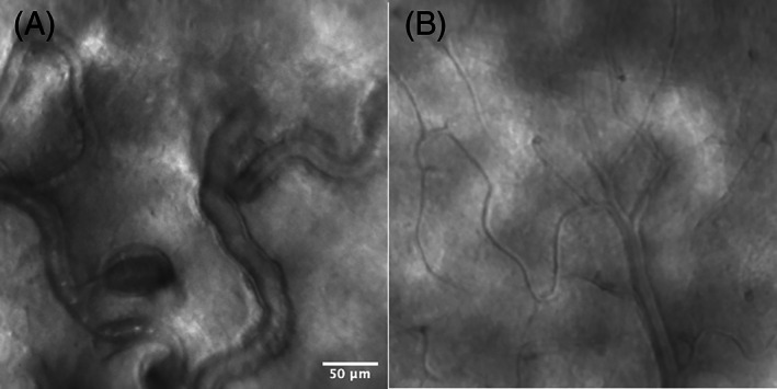
FIGURE 14.

Adaptive optics scanning laser ophthalmoscopy (AO‐SLO) images of a diabetic retinopathy (DR) eye (A) and a healthy eye (B). In the DR eye (A), the capillaries are dilated and beaded with stagnation of blood cells and microaneurysm formation, in contrast to the healthy eye (B) in which the capillaries are of normal calibre and stagnation of blood cell is not observed. Image courtesy of Dr. Shin Kadomoto, Kyoto University Graduate School of Medicine, Kyoto, Japan
