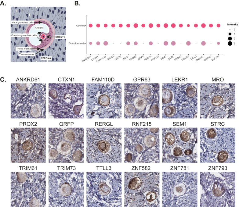
Figure 3.
A schematic overview of ovarian tissue (A). Dotplot summarizing the immunohistochemical staining pattern in oocytes and granulosa cells with the size of dots representing the intensity of the staining (B). Representative images of immunohistochemical stainings of human ovary targeting 18 different MPs (C). The brown color indicates antibody binding. Eleven proteins (ANKRD61, GPR63, LEKR1, PROX2, QRFP, RERGL, RNF215, TRIM61, TTLL3, ZNF781, and ZNF793) showed most prominent protein expression in oocytes, two proteins (FAM110D and TRIM73) showed highest expression in granulosa cells, and five proteins (CTXN1, MRO, SEM1, STRC, and ZNF582) showed equally strong expression in both oocytes and granulosa cells. FAM110D showed cytoplasmic and membranous staining, ZNF781 nuclear staining, SEM1 and RERGL displayed a combination of both cytoplasmic and nuclear staining, and the remaining proteins were exclusively expressed in the cytoplasm.

