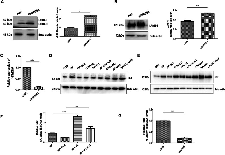
Fig. 4.
HMGB1 inhibition reduces bacterial growth while impairment of lysosomal activity induces bacterial survivability. a–c After transfection for 48 h, nonspecific siRNA (siNS) and HMGB1 siRNA (siHMGB1) transfected cells were further infected with H. pylori SS1 strain (MOI of 100) for 4 h. a, b Immunoblotting was performed in infected cell lysates for expression of LC3B-II and LAMP1. Beta-actin was used as a protein loading control. Densitometry analyses are represented graphically. c Intracellular H. pylori DNA (16SrDNA) was determined by RT- PCR. GAPDH was used as the internal control. Unpaired t-test was performed (d, e) AGS cells incubated with the H. pylori SS1 strain for 4 h and 18 h followed by exposure to glycyrrhizin GLZ (200 µM) and/or chloroquine CQ (50 μM) and/or bafilomycin BAF (50 nM) for 4 h and 18 h treatment respectively. Cell lysates were subjected to a western blot to determine P62 protein levels for 4 h (d) and 18 h (e). Beta-actin was used as a protein loading control. f AGS cells were incubated with the H. pylori SS1 strain for 4 h followed by exposure to glycyrrhizin GLZ (200 µM) and/or chloroquine CQ (50 μM) for 4 h. Intracellular H. pylori DNA (16SrDNA) was measured by RT- PCR. GAPDH was used as the internal control. g Cells were transfected with non-specific siRNA (siNS) and ATG5 siRNA (siATG5) & then incubated with H. pylori SS1 strain for 4 h, and intracellular H. pylori DNA was measured by RT- PCR. GAPDH was kept as an internal control. Graphs were represented as mean ± SEM (n = 3); One-way ANOVA was performed and significance was calculated; **p < 0.01, ***p < 0.001

