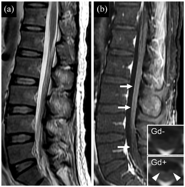
Figure 3.

Representative image of nerve root gadolinium enhancement in patients with autoimmune GFAP astrocytopathy. (a) Sagittal T2-weighted scan shows nerve root disorganization. (b) Sagittal T1-weighted scan with gadolinium shows enhancement alone nerve roots (b; white arrows). Inset in (b) is the axial image before (Gd−) and after (Gd+) gadolinium enhancement, which also shows nerve root enhancement (b; white arrowheads). Gd, gadolinium.
