
Figure 2.
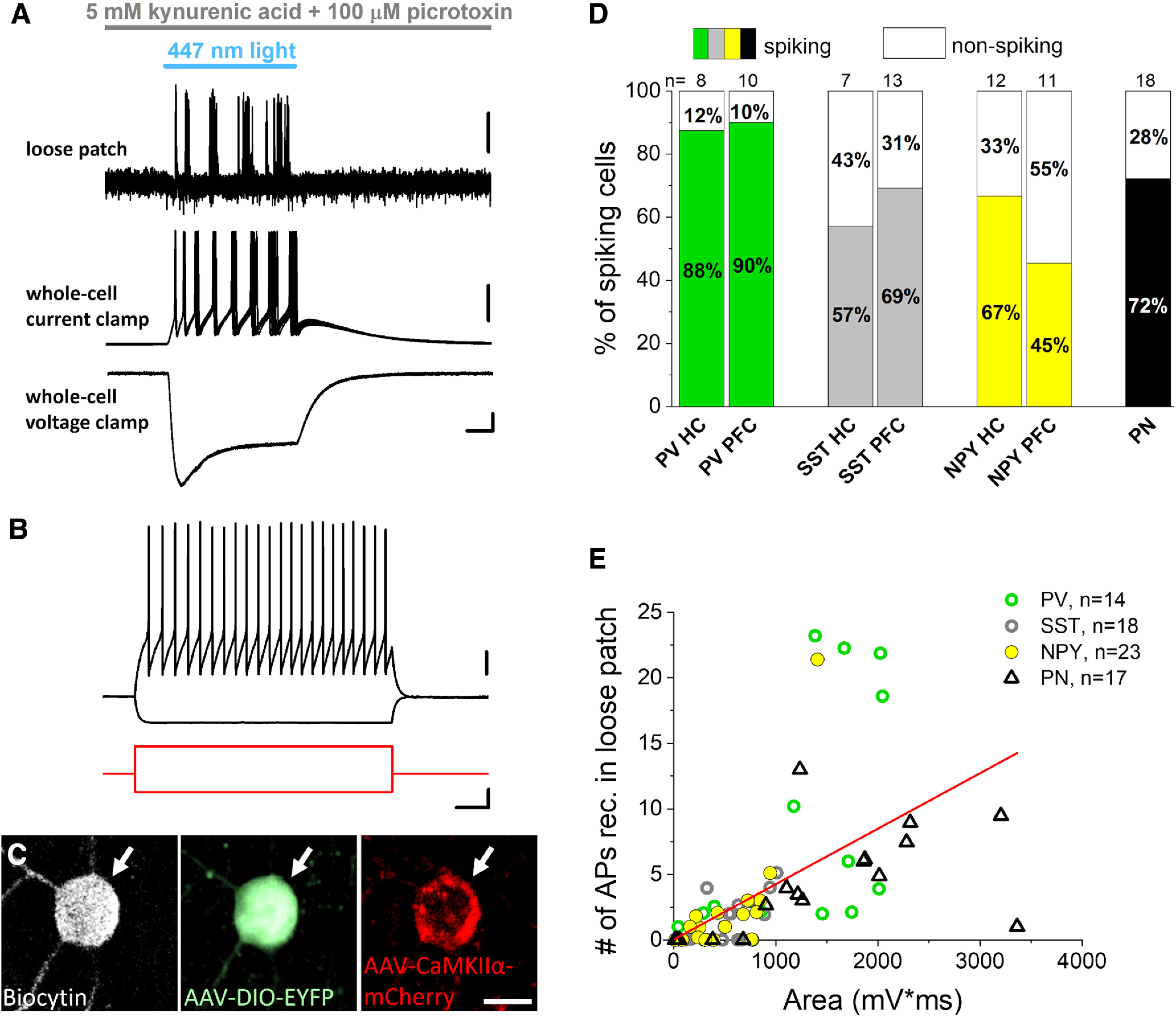
Blue light activation of Channelrhodopsin 2 (ChR2) expressed in parvalbumin (PV), somatostatin (SST), and neuropeptide Y (NPY)-containing interneurons under the control of CaMKIIα promoter readily evokes firing and direct voltage responses. A, Ten consecutive, superimposed voltage traces from loose patch recordings (top) showing action potentials in response to ChR2 activation by blue light illumination in an inhibitory neuron sampled in an acute prefrontal cortical slice prepared from an Npy-Cre mouse. Representative voltage and current responses (middle and bottom) were recorded subsequently in the same interneuron in whole-cell mode as a result of ChR2 activation. Scale bars: loose patch, y = 0.5 mV; current clamp, y = 10 mV; voltage clamp, y = 50 pA and x = 10 ms. B, Voltage responses of the interneuron with fast spiking phenotype (top, black) shown in (A) evoked by two current steps (bottom, red). Scale bars: top, y = 20 mV; bottom, y = 100 pA and x = 100 ms. C, Confocal images demonstrating that the recorded biocytin-filled inhibitory cell (white arrow) same as in A, B expresses Npy promoter-driven EYFP enhanced with anti-GFP immunostaining (green, middle) and CaMKIIα promoter-driven mCherry enhanced with anti-RFP immunostaining (red, right). Scale bar, 10 μm. D, Ratio of spiking neurons on light delivery monitored in loose patch experiments. PN: principal neuron. E, Area of ChR2-evoked voltage responses correlated significantly with the action potential (AP) number detected in loose patch mode during light illumination (Pearson’s r = 0.56, p < 0.001).