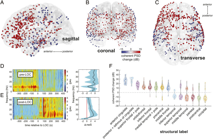
Fig. 2.
Spatiotemporal mapping of preanesthesia and postanesthesia coherent alpha networks. (A–C) Changes in coherent alpha (10-Hz) cPSD across recording sites in all subjects. (D and E) Pre-LOC global coherence principal components depict a narrow 10-Hz rhythm disappearing at LOC (D), and a broader 10-Hz band beginning ~200 s after LOC (E). (F) cPSD is associated with structural (F) parcellations of brain regions. Frontal midline regions such as anterior and posterior cingulate, frontal, and orbitofrontal cortices, as well as the medial temporal lobe, show increased alpha-band cPSD after LOC, whereas posterior regions such as somatosensory and visual areas show a decrease. Constituent labels for each structural category are listed in SI Appendix, Table S3.

