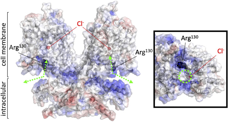
Fig. 3.
An electrostatic potential map of the prestin protein. A ribbon representation of homodimeric human prestin (PDB ID: 7LGU) with surface electrostatic potentials calculated by PyMOL (blue and red indicate positive and negative changes, respectively). The Arg130 residues (black) and bound chlorides (red) are indicated by spheres. Green dashed lines with double arrows indicate presumed anion translocation paths. An intracellular view of one of the protomers is shown in a box on the right. A dashed green circle indicates the intracellular entrance to the anion binding site.

