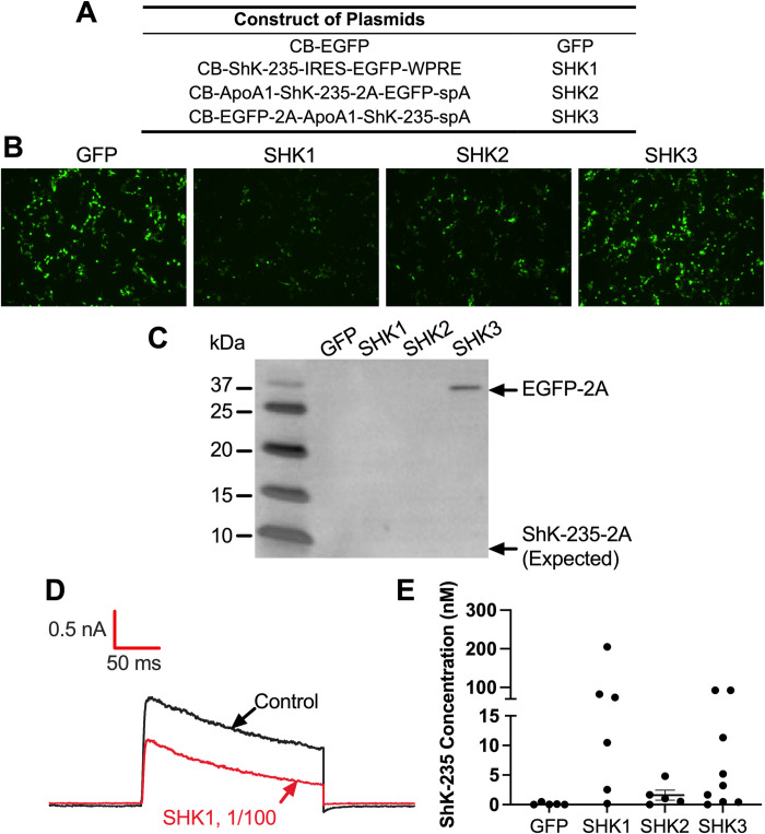
Fig 1. Design and characterization of AAV8-ShK-235 vectors.
(A) Constructs used to express EGFP (GFP) or ShK-235 (SHK1, SHK2, and SHK3). (B) Representative fluorescence microscope images of HEK293T cells 72 hours after transfection with GFP, SHK1, SHK2, or SHK3 plasmids (20 μg of specific plasmid + 60 μg of 1 m/mL of PEI). (C) Western blot showing the detection of the 2A peptide in the supernatants of the HEK293T cells transfected with GFP control, SHK1, SHK2, or SHK3 plasmids, attached to either EGFP (32.7 kDa) or ShK-235 (4.1 kDa). Supernatants were tested without further purification or concentration. (D) Representative whole-cell Kv1.3 currents in L929 fibroblasts stably expressing mKv1.3 before (control) and after application of supernatants from HEK293T cells transfected with GFP control or SHK1, diluted 1:100. (E) ShK-235 concentration in supernatants from HEK293T cells transfected with GFP control, SHK1, SHK2, or SHK3 plasmids. Each data point represents a different recording. The horizontal bar represents the mean concentration.

