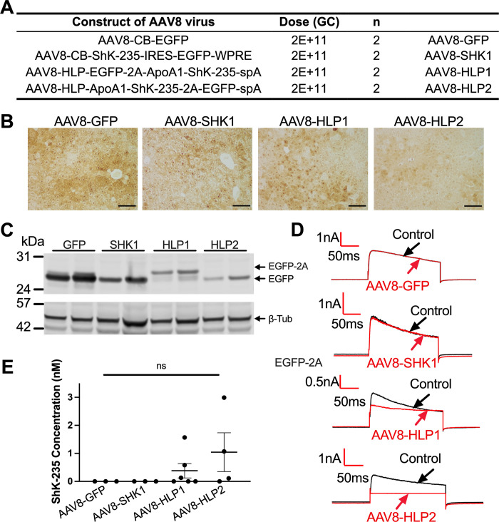
Fig 2. Efficient and specific GFP and ShK-235 peptide expression in mice liver following AAV8-based delivery.
(A) AAV8 constructs and doses injected intraperitoneally into male mice. GC = genome copies. N = 8, or 2 male mice per group. (B) Representative IHC images of mouse livers showing EGFP expression. Scale bars = 100 μm. (C) Western blot analysis of EGFP in liver lysates from mice injected with AAV8 constructs in (A), with β-tubulin (β-Tub) used as a loading control. (D) Representative whole-cell Kv1.3 currents in L929 fibroblasts stably expressing mKv1.3 before (control) and after application of serum from mice injected with AAV8-GFP, AAV8-SHK1, AAV8-HLP1, or AAV8-HLP2, diluted 1:50. Serum was collected 2 weeks post vector delivery. (E) ShK-235 concentration in serum of mice injected with AAV8-GFP, AAV8-SHK1, AAV8-HLP1, or AAV8-HLP2. Each serum sample was tested on 3 cells each.

