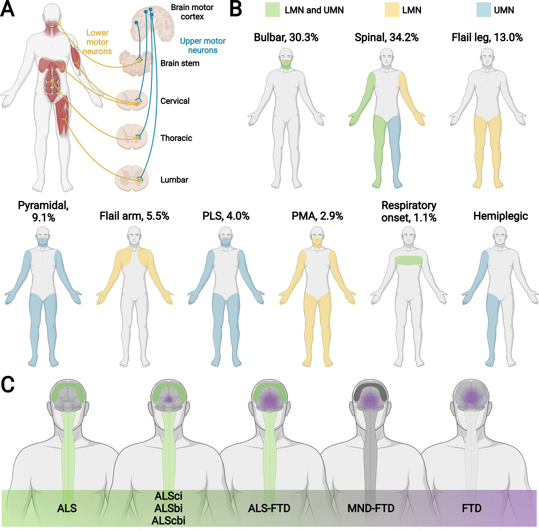
Figure 1. ALS phenotypic variation and spectrum with FTD.

(A) Schematic showing upper motor neurons (UMN; blue), which relay signals from the motor cortex to the lower motor neurons (LMN; yellow, i.e., cranial motor nerve nuclei in the brainstem and anterior horn cells in the spinal cord), which relay signals to the muscles. Motor neurons connecting within the brain stem innervate, among other muscles, cranial muscles. Initial UMN and LMN degeneration in the brain stem are linked to bulbar onset ALS. Motor neurons connecting within the cervical region of the spinal cord innervate, among other muscles, upper limb and respiratory muscles. Motor neurons connecting within the thoracic and lumbar regions of the spinal cord innervate, among other muscles, accessory respiratory, abdominal, and lower limb muscles. Initial UMN and LMN degeneration in the cervical and lumbar regions are linked to spinal onset ALS. (B) ALS patients can present with signs of UMN (blue), LMN (yellow), and combined UMN + LMN (green) dysfunction. Most common ALS phenotypic presentations are bulbar and classical spinal limb onset (cervical, lumbar). Less common ALS phenotypic presentations are flail leg, pyramidal, flail arm, primary lateral sclerosis (PLS), progressive muscular atrophy (PMA), respiratory onset, and hemiplegic. Proportion of various ALS phenotypes shown in the figure as the percentage (%) of a total representative ALS population.(14, 17) Pyramidal is predominantly UMN, as shown in the figure, but still exhibits some LMN signs, differentiating it from PLS. See Appendix Table 1 for more information. (C) ALS occurs on a continuum with FTD. ALS is on one end of the spectrum and presents with pure motor signs from UMN + LMN neurodegeneration (green, spinal cord and motor cortex degeneration). FTD is on the other end of the spectrum and presents with behavioral and/or cognitive deficits from frontotemporal neurodegeneration (purple frontotemporal lobe degeneration). After pure ALS are ALS patients not meeting FTD criteria, defined as ALS cognitive impairment (ALSci), ALS behavioral impairment (ALSbi), and ALS cognitive and behavioral impairment (ALScbi) (green, spinal cord and motor cortex degeneration; small purple sphere of frontotemporal lobe degeneration). Next are ALS patients meeting FTD criteria, defined as ALS-FTD (green, spinal cord and motor cortex degeneration; purple frontotemporal lobe degeneration). Patients on the remainder of the continuum have FTD but do not meet the criteria for ALS. Patients still exhibiting evidence of motor neuron disease (MND) with FTD are defined as MND-FTD (dark grey, spinal cord and motor cortex degeneration; purple frontotemporal lobe degeneration) and patients with no MND signs have FTD (purple, frontotemporal lobe degeneration).
