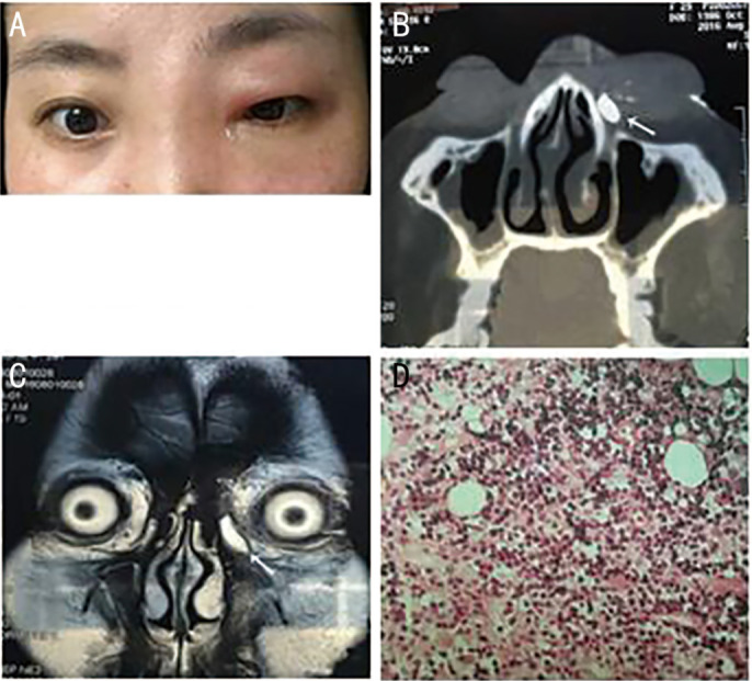
Figure 2. Patient No.8, a 29-year-old female with primary lacrimal sac lymphoma of the left eye (NK/T-cell lymphoma, nasal type) of the left eye.

A: Clinical photograph during early-stage disease, when her symptoms her symptoms suggested acute dacryocystitis. B: Computed tomography dacryocystography (CT-DCG), showing a left post-saccal obstruction, a medial canthus soft tissue mass, and no bone destruction. The arrow indicates the contrast agent (nasolacrimal duct, coronal view). C: Magnetic resonance imaging (MRI), showing the mass had T2 hyperintensity. D: Postoperative pathological examination.
