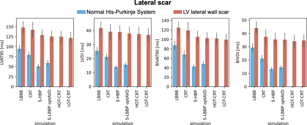
Figure 6. Effect of lateral wall scar on response to pacing.
Bar chart of LVAT-95, LVDI, BIVAT-90 and BIVDI for two different pathologies: proximal LBBB but otherwise normal His-Purkinje system (blue) and scar in the LV lateral wall (orange). The values are presented as mean ± standard deviation (black lines). Abbreviations as in Figure 1.

