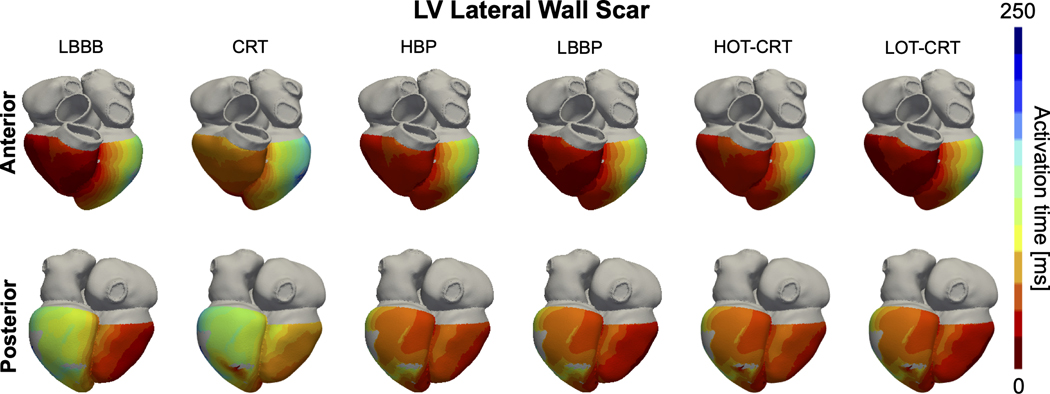
Figure 7. Effect of lateral wall scar on activation times:
Simulated ventricular activation times for one of the patient-specific meshes with proximal LBBB and scar in the LV lateral wall during baseline (LBBB) and pacing. Red and blue areas represent early and late activated regions, respectively. Gray areas in the LV represent scar, simulated as non-conductive tissue. Abbreviations as in Figure 1.

